mast cell tumor cat neck
They reside within many tissues of the body and dogs have a great deal of these cells located within their skin. Head and neck tumors are relatively common in aging cats.

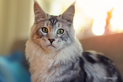
Mast Cell Tumors In Cats
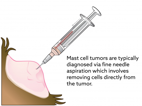
In most instances cutaneous mast cell tumors are diagnosed in cats using a fine needle in order to gather cells to study under a microscope.

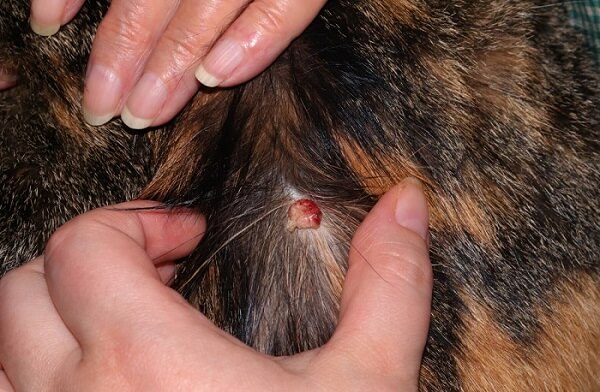
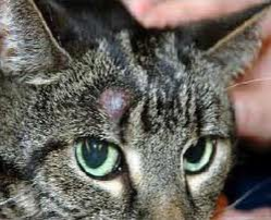
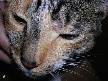
. Typically they are shiny pink hairless nodules on the skin but there are many variations on this. Vet bills can sneak up on you. A mast cell tumor MCT is a type of tumor consisting of mast cells.
Cutaneous visceral and intestinal. Mast Cell Tumor Average Cost. In most instances cutaneous mast cell tumors are diagnosed in cats using a fine needle in order to gather cells to study under a microscope.
Cats affected by these tumors are generally middle-aged and even older. Cutaneous skin mast cell tumours most commonly affect the head and neck and. A mast cell tumor MCT is a tumor that originates from mast cells.
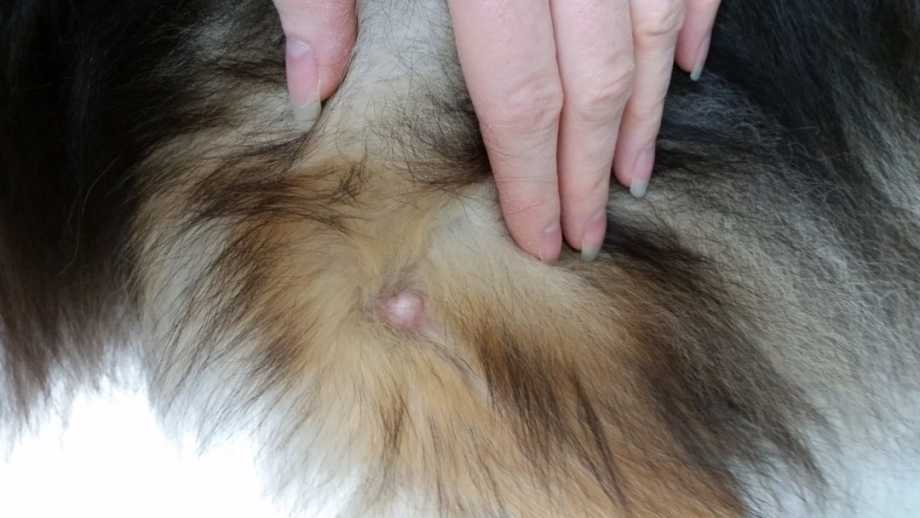
Mast cell tumours commonly affect the skin the spleen in the abdomen andor the intestines. This refers to whether they develop under the skin or on internal tissue respectively. They are small firm raised hairless and can become itchy.
Additionally cutaneous mast cell tumors found on the. Mast cell tumors MCTs are the second most common skin tumor in cats. When signaled by an allergen or the immune system.
Mast cell tumor in cats can occur anywhere on the animals body but is most common on parts such as the skin on the head or neck. While FNA is useful it does not provide any information about the tumors grade level of aggressiveness and biopsy is therefore recommended. How Mast Cell Tumors Affect Your Cat.
Mastocytoma in cats otherwise known as mast cell tumors can be either subcutaneous or visceral. The head and neck regions are the most commonly affected areas especially the top of the head and either or both ears. Diagnosing visceral mast cell tumors can be harder to define.
Mast cell tumors are also sometimes referred to as mastocytomas. They are found primarily in cats older than 4 years old. The head and neck regions are the most commonly affected areas especially the top of the head and either or both ears.
They can be wide plaques or lumps. Some cats will cause self-trauma by itching and chewing during these flare-ups. More Than One Kind of MCT.
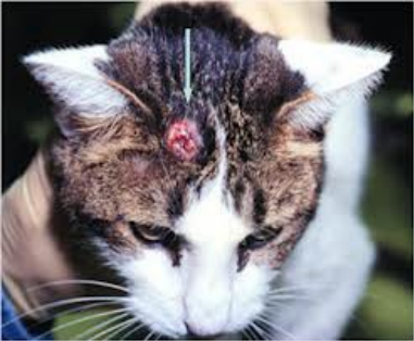
The tumors are single hairless lumps generally 08 to 12 inches 2 to 3 centimeters in diameter. Cutaneous mast cell tumors present as lumps swellings or lesions in the skin or under the skin usually around the head and neck but sometimes elsewhere. 17th August 2018.
Most mast cell tumors are seen as firm plaques hard flattened areas or nodules small lumps in the skin. Chronic Kidney Disease in Dogs and. Visceral or splenic MCTs appear on the spleen or other.
Cholangitis Cholangiohepatitis in Cats. Get the pawfect insurance plan for your pup. Subcutaneous mast cell tumors are more common and are the second most prevalent type of malignant tumor in cats.
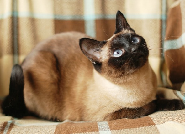
The most common feline breed associated with this disease is the Siamese cat. They are generally found on the cats head and neck but can also affect other parts of the body. The mast cell type is most common.
About 90 of cutaneous MCTs are benign. Mature mast cells contain granules which are basically packets of chemicals. The skin is the most common site for mast cell tumours in cats.
Because these are internal diagnosis is often dependent on the cat owner and their vet noticing changes and behavior and. Understanding the differential diagnoses in this anatomic area is crucial as the diagnostic and therapeutic approaches may vary. Feline mast cell disease is different from mast cell tumors found in canine patients.
Visceral mast cell tumors most frequently affect the. The diagnostic work up in cats is similar to that for dogs. And the remaining 10 are on the head or neck.
Most mast cell tumors arise in the skin but technically they can arise anywhere that mast cells are found. Visceral mast cell tumors occur in the internal organs. Mast cell tumors of internal organs visceral Up to half of all mast cell tumors are visceral and they most commonly affect the spleen.
From 526 quotes ranging from 3000 - 8000. Diagnosing visceral mast cell tumors can be harder to define. Mast cells are immune cells that normally play a role in allergic reactions and inflammatory responses.
In dogs mast cells can develop anywhere and even appear like discolored areas of skin on the nose. Why they mutate is unknown but it is probably due to a variety of factors. It accounts for 8-21 of skin tumors in cats.
In cats about 67 of MCTs also have this mutation. But it must be said that it is quite common for a cat to come out of this pathology in the best conditions including small kittens. What Is a Mast Cell Tumor in Cats.
Cervical Neck Disk Disease in Dogs and Cats. Feline mast cell tumors are commonly found in the head neck and limbs. MCTs can form nodules or masses in the skin and other organs and cause enlargement of the spleen and intestine.
External skin mast cell tumors generally form on the head neck and body but can be anywhere. Mast cell tumors are notoriously invasive and difficult to treat. Even if your cat is not displaying other symptoms.
If your cat has cutaneous mast cell tumors it can recover and lead a normal healthy life after surgery. A mast cell type and a histiocytic type. Unlike the dog the most common locations found in feline patients are the head and neck followed by the extremities.
Additional diagnostic steps will vary depending on the tumors location size and. A mast cell tumor is caused by mutated mast cells. In this case the treatment of choice would be surgical excision.
About half of MCTs affect the visceral organs. What are the signs that my cat may have a mast cell tumor. Mast cells are a type of cell widely distributed in the body and help in the normal immune response.
There are three kinds of MCTs. If the mast cell tumor is injured such as a hard bump during play it can release a large amount of histamine. This lecture will discuss feline oral tumors sinonasal tumors iris melanoma Hodgkins-like lymphoma salivary gland tumors tumors of the ear canal and skin.
In cats mast cells typically develop on the head and neck especially around the base of the ears. Mast cell tumors are initially diagnosed by fine-needle aspriation FNA cytology. Cutaneous MCTs appear on the skin.
They usually protect a cats body from allergens. They may develop anywhere on the body but are most commonly found on the head and neck. The clinical signs of feline mast cell tumors depend on the location of the tumor.
Most mast cell tumors are seen as firm plaques hard flattened areas or nodules small lumps in the skin. Cholangitis and Cholangiohepatitis in Cats. They are the most common splenic tumor second most common skin tumor and third most common intestinal tumor in cats.
Cutaneous mast cell tumors affect the skin and account for approximately 20 of skin tumors in cats. Two distinct variants occur. The skin form of the feline mast cell tumor typically arises around the head and neck and lesions may be solitary or multiple.
What are the signs that my cat may have a mast cell tumor. What does a mast cell tumor look like on a cat.
Mast Cell Tumors In Cats
Figure 3 From Mast Cell Tumors In Cats Semantic Scholar
Skin Tumours Histiocytoma In Cats Causes Symptoms Treatment All About Cats
2
Mast Cell Tumors Veterian Key

Diagnosis And Treatment Of A Feline Oral Mast Cell Tumor Sciencedirect
Mast Cell Tumor Mastocytoma In Cats Petmd

Feline Mast Cell Tumours Blackwood 2015 In Practice Wiley Online Library

Mast Cell Tumors In Cats Symptoms Diagnosis Treatment All About Cats

Mast Cell Tumors In Cats Symptoms Causes And Treatments
Mast Cell Tumors In Cats Vca Animal Hospitals
Mast Cell Tumors Veterinarian In Montgomery Al Animal Hospital Of Montgomery
Mast Cell Tumors Veterian Key
Mast Cell Tumors Mastocytomas In Dogs Small Door Veterinary

Feline Mast Cell Tumours Blackwood 2015 In Practice Wiley Online Library

Feline Mast Cell Tumours Blackwood 2015 In Practice Wiley Online Library
Mast Cell Tumors Veterinarian In Montgomery Al Animal Hospital Of Montgomery

Figure 3 From Mast Cell Tumors In Cats Semantic Scholar

Animal Surgical Center Of Michigan Veterinarian In Flint Mi